Congratulations to Hélène Roberge at the physics of materials and nanostructures group of the Institute of Materials Jean Rouxel (IMN) of Nantes and the Process engineering for environment and food (GEPEA) laboratory in Saint Nazaire on receiving the 1st prize in the RIME 2020 poster competition.
The RIME (Réseau d’imagerie en Microscopie électronique – Electron Microscopy Imaging Network) is a national electron microscopy specialist network (≈ 300 members).
The network currently brings together joint centres and laboratories focused on the scientific and technical development of electron microscopy in biology. It has 300 members belonging to different supervisory bodies (Universities, CNRS, INSERM, INRA) spread over several French sites.
It is now called RIME, the Electron Microscopy Imaging Network.
This competition is open to all RIME members present at these annual days. It is sponsored by the company » LFG distribution / DiATOME « .
The posters will be exhibited throughout the days and will be the subject of a short presentation: « my poster in 120s », a « poster evening » and a vote by the participants.
The first Poster prize will be rewarded with a jewellery diamond awarded by the company « LFG Distribution / DiATOME ».
Transmission or Scanning Electron Microscopy Photo Competition Organised by RIME on the occasion of the Annual Meetings, from 11 to 13 May 2020 in Grenoble.
This competition is sponsored by the company DELTA Microscopies. It is open to all RIME members present at these annual days.
The first six photos will be rewarded with prizes awarded by the company Delta Microscopies (one prize per person). The selected photos will be published in the 2021 calendar published by Delta Microscopies.
Every year, a workshop-style conference is organized with approximately 100 people in one center of microscopy. The goal of this days is to share tips and technic, meet other specialist and facilitate exchanges about problematics and research in biology/sciences electron microscopy imaging and spectroscopy. This year, it was on Autrans – Grenoble with The ibs lab (Institut de biologie structural – Institute of Structural Biology, CNRS / CEA / University of Grenoble) between the 30/09 and the 02/10/2020 on the theme « Characterization of nano-objects in electron microscopy ».
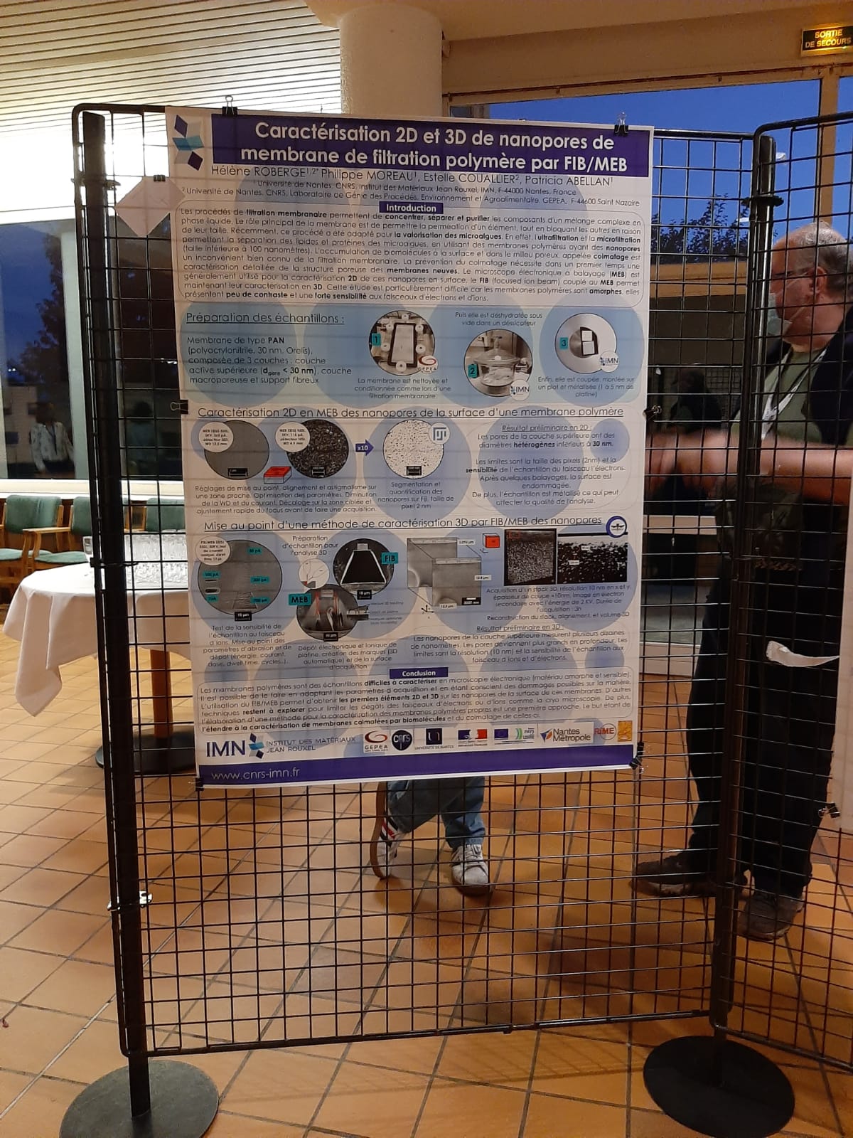
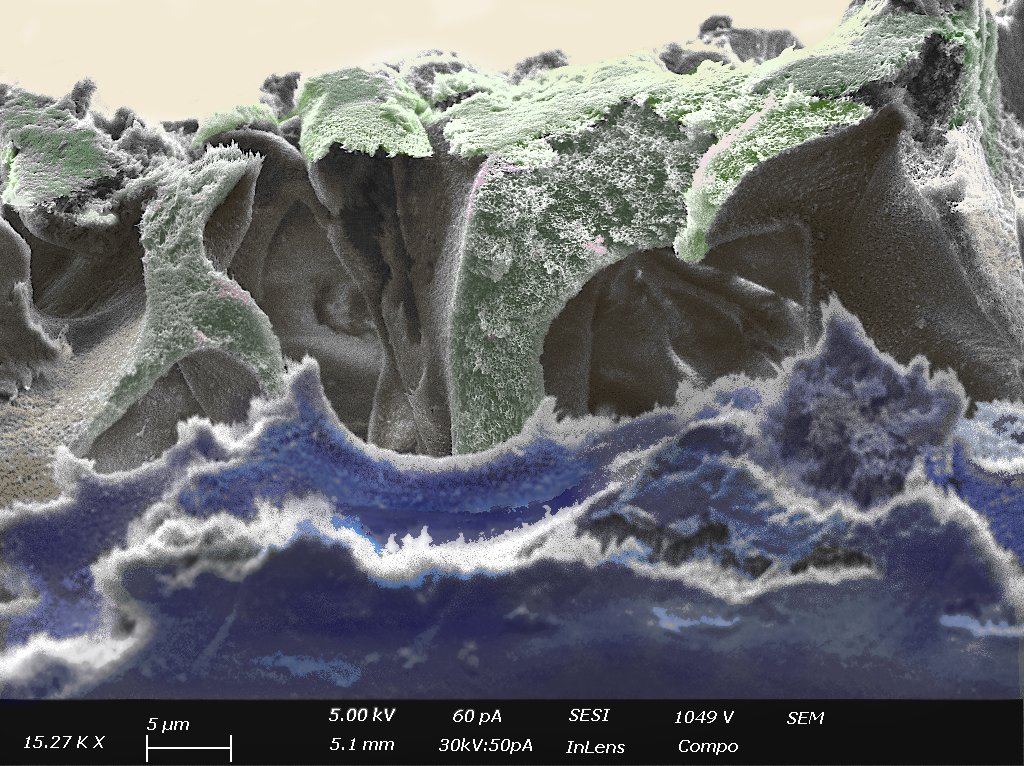
It’s poster was based on her first year Phd work with the FIB/SEM technic development on high resolution characterization within sensitive polymer membrane, titled “2D and 3D characterization of polymer filtration membrane nanopores by FIB/MEB”.

About Hélène Roberge,
Phd student in IMN Nantes – Institute of Material Jean Rouxel and in GEPEA Saint Nazaire – Laboratory of Process Engineering, Environment and Food Processing , supervised by Patricia Abellan (IMN), Estelle Couallier (GEPEA) and Philippe Moreau (IMN).
Specialized in electron microscopy, she works on polymer filtration membrane characterization. Ultrafiltration and microfiltration have been adapted for microalgae valorization, for which porous polymer membranes are used. The accumulation of biomolecules at the surface and in the porous medium, termed fouling, is a major operational challenge and a well-known drawback in membrane filtration. The prevention of fouling requires a detailed characterization of the structure of clean filtration membranes as well as their interaction with the different target biomolecules. She uses FIB/SEM to image (2D et 3D) nanopore on the surface and detailed structure of clean membrane in the first place. Which are challenging due to the sensitivity and the less contrast of the sample.

